Jonathan Sheard, Ph.D., Sheard BioTech Ltd & University of Reading, United Kingdom
Introduction
Mesenchymal stem cells (MSCs) are clinically relevant, tripotent adult stem cells that give rise to bone, cartilage and fat cells. MSCs can be efficiently isolated from various sources including bone marrow, fat and many dental tissues. In vitro, MSCs can be cultivated as conventional 2D monolayer cultures and, as reported recently [1], within nanofibrillar cellulose (GrowDex®). It has also been suggested, that cultivation of MSCs as 3D spheroids might increase their anti-inflammatory potential [2,3]. Briefly, MSCs cultured within hanging drops begin to aggregate and form spheres within 3 days [4] and these spheres remain viable. However, the number of apoptotic or necrotic cells may increase relative to a higher seeding density of cells or culture time [2]. Additionally, MSCs cultured as spheroids show higher differentiation potential towards catilage regeneration when cultured within 3D scaffolds or as hanging drop spheres [5,6].
This application note describes an easy method for generation of MSC-spheres using GrowDex®. Utilising a 3D matrix such as GrowDex in the hanging drop technology may provide a useful tool for examining; cell-cell and cell-matrix behaviour , in addition to cell differentiation potential, within smaller sample volumes and with fewer cells.
Materials
- Adipose Derived Mesenchymal Stem Cells (ADMSCs, Cat# PT-5006, Lonza)
- Complete Media: DMEM (High Glucose) supplemented with 10% FBS, 1% Pen/Strep and 1% L-Glutamine (Sigma Aldrich)
- Non-treated tissue culture dishes - 60x15mm (Cat# 83.3901.500, Sarstedt)
- GrowDex, 1.5% (Cat# 100 103 005, UPM)
- EVOS Live imaging system (ThermoFisher)
- Image-J image analysis software (NIH)
Method
- ADMSCs were cultured in complete media and incubated at 37°C with 10% CO2.
- Following trypsinization, cells were resuspended in media at a concentration of 1 x 10⁶cells/ml.
- Subsequently, cells were mixed with the appropriate volume of media and GrowDex to provide final concerntrations of 0.2% and 0.4% GrowDex seeded with 100 cells/µl.
- A droplet of 10 µl per sample was pipetted onto the lid of a non-tissue culture treated petri-dish. No less than 3 droplets were prepared per sample.
- Petri-dishes were incubated at 37°C with 10% CO2.
- 5 µl of complete culture media was added every 1 to 2 days.
- Images were taken using the EVOS imaging system using 4x and 10x objectives. Images captured using the 10x objective were loaded into image J where the number and area of the cell spheres was measured and quantified.
Results
Through 7 days of culture expansion, ADMSC spheres were visible within all hanging drop samples (Fig.1). The spheres varied in both size and number depending on the the concentration of GrowDex used for the culture (Fig.2).
Small spheres were observed in all hanging drops irrespective of the GrowDex concentration used (Fig.1). The number and size of spheres was quantified for each hanging drop (Fig.2). At 7 days of culture, GrowDex was observed to condense within the hanging drop (Fig.1 C, G). It was also noted, at the lower concentration of 0.2% (Fig.1 C), GrowDex did not maintain a regular pellet shape compared to that of the higher concentration of 0.4% (Fig.1 G).
At day 1, an average of 55 cell spheres were counted in 0.2% GrowDex hanging drops (Fig.1 A-B, Fig.2 A), with an area of 124 µm2 per sphere (Fig.2 B). After 7 days of culture, the number of spheres increased approximately 2 fold to 116 (Fig 2. A) and the area also increased approximately 2 fold to 250 µm2 per sphere (Fig.2 B).
In 0.4% GrowDex an average of 38 cell spheres per hanging drop where counted on day 1 which increased to 77 spheres per hanging drop by day 7 (Fig.2 A). The area of each sphere at day 1 was approximately 133 µm2, increasing only 1.5 fold to 203 µm2 per cell sphere.
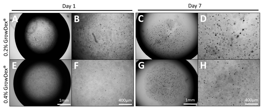
Fig 1. Images showing primary ADMSCs cultured as hanging drops through 7 days using GrowDex concentrations of 0.2% (A-D) and 0.4% (E-H). Images were captured at 4x (A, C, E, G) and 10x (B, D, F, H) magnification.

Fig 2. Quantification of the average number of cell spheres per hanging drop (A) and the area (µm2) per sphere from images collected of droplets at Day 1 (B) and Day 7 (C).
Conclusions
Embedding cells within a 3D matrix and culturing these as hanging drops, enables the observation and measurement of both spatial cell-cell interactions and cell-matrix interactions within a small volume [7]. Combining multiple cell types as a co-culture system can also provide further opportunity to closely mimic in vivo conditions [8]. Additionally, retrieval of the spheres from hanging drop cultures has been detailed and is quite simple [4].
MSC spheres cultured within hanging drops have been shown to posses enhanced osteogenic and adipogenic differentiation potential [9], and produce higher amounts of cartilage matrix deposition [6]. Moreover, conditioned media harvested from MSCs spheres has been shown to have stong anti-inflammatory effects [2, 3, 4].
Here, an alternative method of culturing MSC spheres within GrowDex has been demonstrated. The results show that MSCs can be easily embedded in GrowDex and, following 7 days of culture, these MSCs form small compact cell spheres which increase in size and number. This method offers a useful alternative approach to study cell behavioiur, interaction and differentiation potential, with the added benefit of requiring smaller sample volumes and fewer cells.
References
- Azoidis, I., J., et al. (2017). "Three-dimensional cell culture of human mesenchymal stem cells in nanofibrillar cellulose hydrogels." MRS Communications: 1-8.
- Bartosh, T. J., et al. (2010). "Aggregation of human mesenchymal stromal cells (MSCs) into 3D spheroids enhances their anti-inflammatory properties." Proceedings of the National Academy of Sciences of the United States of America 107(31): 13724-13729.
- Ylöstalo, J. H., et al. (2012). "Human mesenchymal stem/stromal cells (hMSCs) cultured as spheroids are self-activated to produce prostaglandin E2 (PGE2) that directs stimulated macrophages into an anti-inflammatory phenotype." Stem cells (Dayton, Ohio) 30(10): 2283-2296.
- Bartosh, T. J. and J. H. Ylostalo (2014). "Preparation of anti-inflammatory mesenchymal stem/precursor cells (MSCs) through sphere formation using hanging drop culture technique." Current protocols in stem cell biology 28: Unit-2B.6.
- Wang, L., et al. (2009). "Effect of Initial Seeding Density on Human Umbilical Cord Mesenchymal Stromal Cells for Fibrocartilage Tissue Engineering." Tissue Engineering. Part A 15(5): 1009-1017.
- Suzuki, S., et al. (2012). "Properties and usefulness of aggregates of synovial mesenchymal stem cells as a source for cartilage regeneration." Arthritis Research & Therapy 14(3): R136-R136.
- Matak, D., et al. (2017). "Colony, hanging drop, and methylcellulose three dimensional hypoxic growth optimization of renal cell carcinoma cell lines." Cytotechnology 69(4): 565-578.
- Edmondson, R., et al. (2014). "Three-Dimensional Cell Culture Systems and Their Applications in Drug Discovery and Cell-Based Biosensors." Assay and Drug Development Technologies 12(4): 207-218.
- Frith, J. E., et al. (2010). "Dynamic three-dimensional culture methods enhance mesenchymal stem cell properties and increase therapeutic potential." Tissue Eng Part C Methods 16(4): 735-749.