Jeroen Pouwels¹, Topi Tervonen¹, Jonathan Sheard², Piia Mikkonen²
1. Finnish Genome Editing Center-Helsinki (FinGEEC-Helsinki), University of Helsinki, and Biocenter Finland, Finland.
2. UPM Biomedicals, Helsinki, Finland
INTRODUCTION
Despite the discovery of many new treatment modalities for cancer, most patients experience little to no long-term benefits due to the lack of predictive biomarkers [1]. Therefore, a great unmet medical need exists for personalized assays, for example robust patient-derived 3D cancer culture assays to support clinical decision making or for genome editing and drug discovery purposes.
For such personalized medicine solutions to become translational, it is imperative that patientderived material can be cultured ex vivo to provide a clean and truly representative snapshot of the tumor microenvironment. This can be done using the patient-derived explant culture (PDEC) method. However, this has commonly been carried out in animal derived matrices, leading to changes in the tumor microenvironment, cell behavior, proliferation as well as differentiation, which are all influenced by matrix derived growth factors and extracellular matrix molecules [2].
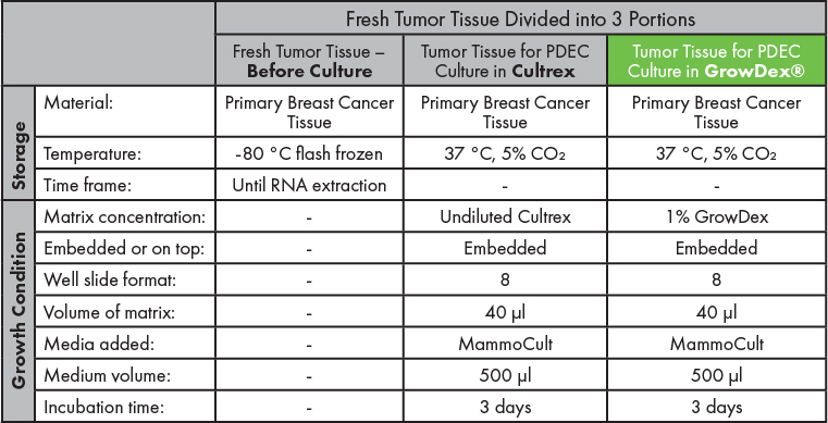
As illustrated graphically in Figure 1, the suitability of RNA isolation, purification and concentration was assessed from fresh breast cancer tumor tissue (Figure 1a), and similar tissue sections cultured as PDECs in GrowDex diluted to 1.0% (Figure 1b) or in Cultrex at 100% (Figure 1c). Readouts of RNA quality and quantity are presented here.

Figure 1. Schematic representation of the experimental layout and analysis of RNA isolated from fresh tumor tissue ‘before culture’ (a) or following PDEC culture in either GrowDex diluted to 1.0% (b) or Cultrex at 100% (c).
MATERIALS
- GrowDex (Cat No. 100 103 005, UPM Biomedicals)
- Cultrex (Cat No. 3533-005-02, R&D Systems)
- MammoCult growth media (Cat No. 05620, StemCell Technologies)
- MammoCult proliferation supplements (Cat No. 05621, StemCell Technologies)
- Collagenase A (Cat No. 10103578001, Sigma-Aldrich)
- Heparin (Cat No. E4643, Sigma-Aldrich)
- Gentamicin (Cat No. 345814-M, Sigma-Aldrich)
- Penicillin/Streptomycin (Cat No. P4333, Sigma-Aldrich)
- 8-well chamber slides (Cat No. 177402, Thermo Fisher Scientific)
- TRIzol (Cat No. 15596026, Invitrogen)
- Precellys® Ceramic kit, 0,1 mm, 50×2 ml tubes, pre-filled with ceramic beads (Cat No. 10144-500, Bertin)
- NanoDrop™ One/OneC Microvolume UV-Vis Spectrophotometer (Thermofisher)
- Qubit™ RNA HS Assay Kit (Cat No. Q32852, Invitrogen)
- 4200 TapeStation System (Cat No. G2991BA, Agilent)
- NextSeq 500 System WGS Solution (Illumina)
- RNA clean & concentrator kit (Cat No. R1017, Zymo Research)
METHODS
1. Tumor tissue collection and processing
Primary breast cancer tissue from consenting patients (N=4), approved by the Hospital District of Helsinki and Uusimaa, (HUS) was collected from the HUS pathology department. Tumor tissue was kept on ice during transportation between hospital departments.
In the lab, the tumor was divided into 3 portions. One portion of the fresh tumor tissue sample was set aside and temporarily stored by flash freezing at -80 °C prior to RNA extraction and RNA sequencing (Figure 1a). This sample is titled as ‘before culture’. The other two similar-sized tumor tissue portions were processed for PDEC culture in either GrowDex or Cultrex prior to RNA extraction and RNA sequencing (Figure 1b and c respectively). Summarized in Table 1.
2. PDEC cultures
PDEC cultures were prepared as detailed in Munne, P.M. et al., (2021). Briefly, tumor material was processed into small fragments through incubation overnight (O/N) with gentle shaking (130rpm) at 37°C in MammoCult basal medium containing 0.2% of Collagenase A, MammoCult proliferation supplements, 4μg/ml heparin, 50μg/ml gentamicin, and penicillin/streptomycin (diluted 1:100).
The next day the mixture was centrifuged at 1300rpm for 3min and the pellet was resuspended in 1ml MammoCult media. Fragments were recentrifuged, resuspended in 1.0% GrowDex or undiluted Cultrex and seeded to 8-well chamber slides for 3D culture.
GrowDex was prepared by diluting the stock 1.5% to the desired concentration of 1.0% according to the manufacturer’s instructions. Briefly, 500 µl of MammoCult media was placed into an Eppendorf tube, then 1 ml of GrowDex was added. This was mixed thoroughly and used as necessary.
In total, 40 μl of matrix was added per well in the 8-well chamber slides and each well was supplemented with 500 μl of MammoCult growth media. PDEC cultures were incubated in a standard cell culture incubator (37 °C, 5% CO₂) for 3 days. Summarized in Table 1.
3. RNA isolation and purification
Total RNA was isolated with TRIzol™ and RNA was purified with the Zymo RNA Clean & Concentrator™ kit. RNA isolation from fresh tumor tissue is described below in (3.a) and RNA isolation from PDEC cultures is described in (3.b).
-
- RNA isolation from fresh tissue
After the fresh tumor tissue sample had been temporarily stored by flash freezing at -80°C, it was thawed and placed in 1 ml TRIzol™ reagent. Tumor pieces were lysed using Precellys ceramic beads according to the manufacturer’s instructions. - RNA isolation from PDECs
PDEC samples were snap frozen after culturing and stored at -80 °C, after which the samples were thawed and mixed with 1ml TRIzol™ reagent and subsequently lysed by passing the samples through 20G needles.
- RNA isolation from fresh tissue
The isolated RNA samples detailed above in 3.a and 3.b, were then incubated for 5 minutes to allow complete dissociation of the nucleoprotein complexes. After 0.2 ml of chloroform was added per 1 ml of TRIzol™, the samples were mixed thoroughly by shaking, followed by an incubation of 2-3 minutes. Subsequently, the samples were centrifuged for 15 minutes at 12,000 × g at 4 °C. The mixture separates into 3 phases: a lower red phenol-chloroform phase, an interphase, and a colorless upper aqueous phase. The aqueous phase containing the RNA was transferred to a new Eppendorf tube by angling the tube at 45 ° and pipetting the solution out.
RNA preps were purified and concentrated immediately after extraction with the Zymo RNA Clean & Concentrator™ kit according to the manufacturer’s instructions. RNA was eluted in RNAse-free water and the concentration was measured with Nanodrop. RNA was frozen at -80 °C until RNA sequencing.
4. RNA analysis
The Biomedicum Functional Genomics Unit at the Helsinki Institute of Life Science and Biocenter Finland at the University of Helsinki performed the following assays as a service: RNA concentration measurements using a Qubit™ RNA HS Assay Kit, RNA quality confirmation using a TapeStation system and bulk mRNA sequencing. All RNA quality analysis was done using the Agilent Tapestation 4200: High Sensitivity RNA ScreenTape, according to manufacturer’s instructions. RNA concentrations were measured using Qubit (Thermo Fisher), according to manufacturer’s instructions.
RESULTS
The experiments described above and depicted in Figure 1 were performed using primary breast cancer tissue collected from consenting patients (4 patients in total). Each tumor tissue sample was successfully divided into 3 portions for RNA analysis before culture or following culture as PDECs in either Cultrex or GrowDex. Immunofluorescence imaging of PDECs can be seen in Figure 2, following 7 days of culture in an animal derived matrix (Figure 2 left panel) or in GrowDex (Figure 2 right panel) [4]. RNA isolation, purification and concentration were performed on all samples as detailed above.
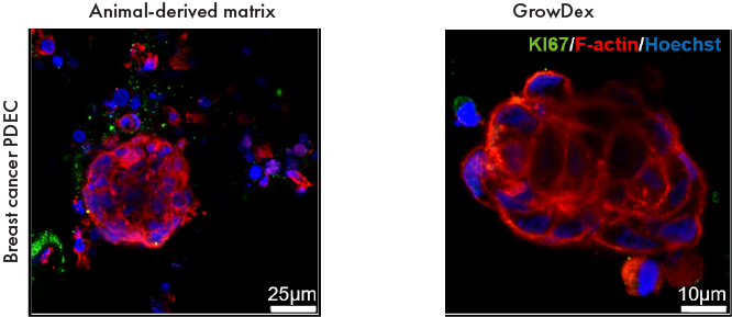
Figure 2. Immunofluorescence images of primary breast cancer tissue samples stained with KI67 (green), F-actin (red) and Hoechst (blue) following 7 days culture in an animal-derived matrix (left panel) or GrowDex (right panel).
High quality RNA was successfully derived for RNAseq (Figure 3) from fresh tumor tissue before culture, or tissue cultured as PDECs in either Cultrex or GrowDex. The quality of RNA is shown by the RNA Integrity Number (RIN value) reported by the Agilent Tapestation 4200. It can be seen that the quality of RNA isolated from tissue cultured as PDECs in GrowDex was considerably greater than the RNA isolated from similar PDECs in Cultrex or from fresh tissue samples before culture.
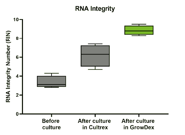
Figure 3. RNA Integrity Number (RIN) values of RNA isolated from tumour tissue samples before culture, or after PDEC culture in Cultrex or GrowDex. Data shown is N=4 with min to max.
As a part of the RNA analysis, the library size was measured. The library size is the number of sequenced reads mapping to the reference genome for each sample. This data is shown in Figure 4 where the quantity of isolated RNA from PDECs in GrowDex were comparative to the quantity of RNA isolated from fresh tissue, these were both greater than the quantity of RNA isolated from PDECs in Cultrex.
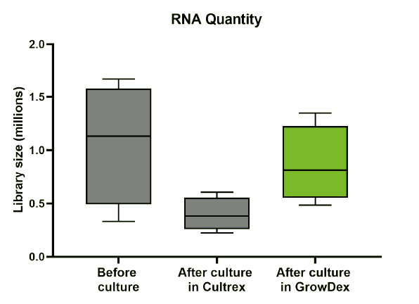
Figure 4. Library size of tumour tissue samples before culture, or after PDEC culture in Cultrex or GrowDex. Data shown is N=4 with min to max.
CONCLUSIONS
This study clearly demonstrates that high quality RNA can be isolated from cells and tissue cultured as PDECs in GrowDex. The quality of this RNA is greater than that isolated from PDECs cultured in Cultrex or even freshly isolated tissue. Additionally, the quantity of isolated RNA, i.e., the library size, from PDECs in GrowDex were comparative to fresh tissue, and greater than that isolated from PDECs in Cultrex.
RNA mirrors the sequence of the DNA from which it was transcribed. High quality and quantity of isolated RNA enables the study and analysis of a cell transcriptome-wide differential gene expression and differential splicing of mRNAs. Specifically, by analyzing an entire collection of RNA sequences in a cell (i.e., the transcriptome), researchers can determine when and where each gene is turned on or off in the cells and tissues. Furthermore, understanding gene expression analysis is especially important during ex vivo drug screening, allowing researchers to see how the tumor sample responds to the different drug treatments.
REFERENCES
- Sharma, P. and Allison, James P. (2015). "Immune Checkpoint Targeting in Cancer Therapy: Toward Combination Strategies with Curative Potential." Cell 161(2): p. 205-214.
- Yue, B. (2014). "Biology of the Extracellular Matrix: An Overview." Journal of Glaucoma 23.
- Munne, P.M., et al. (2021). "Compressive stress-mediated p38 activation required for ERα+phenotype in breast cancer." Nature Communications 12(1): p. 6967.
- Munne, P. "TIME - Tumor Immune MicroEnvironment." Speaker Presentation at UPM Annual Conference 2020.